Research - (2022) Volume 5, Issue 4
Anterior Segment Disorders and Dry Eye Injuries, Allergens, Inflammation, Dry Eye, Corneal Disorders, Cataract and Presbyopia Can Affect the Eye's Ability to Focus
Ahmadull Bari1* and AK Al-Miraj22Research Assistant, Department of Vascular Surgery, Bangabandhu Sheikh Mujib Medical University,, Dhaka, Bangladesh
Received: Nov 10, 2022, Manuscript No. AMDHS-22-79474; Editor assigned: Nov 12, 2022, Pre QC No. AMDHS-22-79474 (PQ); Reviewed: Nov 15, 2022, QC No. AMDHS-22-79474 (Q); Revised: Nov 17, 2022, Manuscript No. AMDHS-22-79474 (R); Published: Dec 02, 2022, DOI: 10.5530/amdhs.2022.4.11
Abstract
Background and objectives: Dry Eye is a common condition that is often under diagnosed. Normal vision requires moist healthy ocular surface. A sufficient quality of tears, normal composition of tears film, lid closure to maintain healthy ocular surface. Dry eye is a disorder characterised by either quantitative decrease or qualitative change in pre-corneal film resulting in spectrum. Along with assessment of dry eye with OSDI questionnaire and various tests were done to grade dry eye.
Methods: A prospective study was conducted to assess the dry eye in July 2021 to August 2022 in BSM Medical University Dhaka and Dishari Eye Hospital Chattogram, Bangladesh. Total 63 cases were chosen from the outpatient department of and assessment of dry eye was made by tests like Schirmer’s test, Tear breakup time, Rose Bengal dye test. Ocular Surface Disease Index (OSDI) questionnaire was given to patients to assess the grading of dry eye.
Results: Majority 31.7% patients were in the group 41 years to 50 years. In the study OSDI questionnaire had a good reliability and consistency (p< 0.001) highly significant. Pearson correlation with r value among various test like Rose Bengal test, Schirmer’s test and TBUT showed a good correlation.
Conclusion: Dry eye is a chronic disease and increase in incidence of dry eye increases with age. Various risk factors should be assessed to find the cause of dry eye. Subjective test like Odi questionnaire with objective tests like rose Bengal, Schirmer’s and Tear breakup time should be done routinely to assess the grading of dry eye so that long term complications associated can be prevented.
Keywords
OSDI, Schirmer’s, Rose Bengal, Tear break up time
Introduction
Dry Eye is a common condition that is often under diagnosed. Normal vision requires moist healthy ocular surface. A sufficient quality of tears, normal composition of tears film, lid closure to maintain healthy ocular surface [1]. Dry eye is a disorder characterised by either quantitative decrease or qualitative change in pre-corneal film resulting in spectrum of pathological changes that may adversely affect the ocular surface resulting in ocular surface disorders often leading to conjunctival squamous metaplasia and punctate epithelial erosion of cornea [2, 3]. Dry eye results in discomfort and visual disturbance and tear film Instability with potential damage to ocular epithelial surface and accompanied by increase in tear osmolarity and inflammation. Dry eye syndrome involves multiple risk factors that when disregarded can result in treatment failure and frustration both for the patients and the physician. Dry eye may lead to increased risk of infections, medications toxicity, contact lens intolerance, progressive ocular surface disease, scarring, cornea morbidity namely keratinisation, corneal thinning, vascularisation, microbial and sterile corneal ulcer leading to perforation and severe visual loss. Hence correct diagnosis and appropriate management of dry eye is essential [4]. Dry eye etiology is not a single disease entity. It is a disorder of lacrimal function unit according to some. In recent population based surveys it indicates that dry eye affects millions of people in worldwide as many as 20% to 25% patients reported to outpatient department complaints of dry eye symptoms making it very common presentation. It poses a huge economic burden to patients if not evaluated for risk factors, etiopathogenesis that may result in delay in treatment. It affects the quality of life of patients with regards to daily visual acuity. It results in social stigma as a patient may have chronic red eye. Patient may undergo into depression which requires additional psychotherapy. The purpose of approach to is to assess dry eye with OSDI questionnairre along with battery of tests so as to improve patients comfort and prevent structural damage to ocular component. Due to lack of uniformity in definition and inability of any single diagnostic test or set of diagnostic test to confirm or rule out the condition [5].There has been a shift towards symptom based assessment as a key component in clinical diagnosis with grading of severity of dry eye. Use of symptom based validated questionnaire might be beneficial as it allows grading of symptoms and is repeatable for comparative purpose before, during and after treatment. Recent advances in treatment suggests the use of lubricants, anti-inflammatory drugs, plugs to augment the tear film [6].
Materials and Methods
A prospective study was conducted to assess the dry eye in July 2021 to August 2022 in BSM Medical University Dhaka and Dishari Eye Hospital Chattogram, Bangladesh. Total 63 cases were chosen from the outpatient department of and assessment of dry eye was made by tests like Schirmer’s test, Along with assessment of dry eye with OSDI questionnaire and various tests were done to grade dry eye. The material for the study was collected from the patients presenting themselves directly to Department of Ophthalmology.
■Inclusion criteria
Both male and female patients equal to and above 20 years presenting with fallowing symptoms Burning sensation, sandy grity feeling, Foreign body sensation, Photophobia, Heavy lids. The above symptoms increase in conditions of low humidity and wind.
■ Exclusion criteria
1 Patients less than 20 years of age, Patients with H/O increased mucoid discharge and watery secretion suggestive of vernal keratoconjunctivitis, Patients with H/O Alkali burns, Patients with H/O Trachoma, Patients with H/O Acute ocular infections, Patients with H/O Ocular surgery within last 6 months, Patients with H/O Impaired eyelid function like in Bells palsy, nocturnal lagophthalmos, ectropion and Contact lens users.
An OSDI-Allergan ocular surface disease index questionnaire is to be administered to all participants to assess the symptoms of dry eye and correlate them with OSDI (provided by Allergan, Inc. Irvine, CA) was used to quantify the specific impact of dry eye [7]. This disease-specific questionnaire includes three subscales: ocular discomfort (OSDIsymptoms), which includes symptoms such as gritty or painful eyes; functioning (OSDI- function), which measures limitation in performance of common activities such as reading and working on a computer; and environmental triggers (OSDI-triggers), which measures the impact of environmental triggers, such as wind or drafts, on dry eye symptoms. The questions are asked with reference to a one-week recall period. Possible responses refer to the frequency of the disturbance: none of the time, some of the time, half of the time, most of the time, or all of the time. Responses to the OSDI were scored using the methods described by the authors. Subscale scores were computed for OSDI-symptoms, OSDI-function, and OSDI-triggers, as well as an overall averaged score. OSDI subscale scores can range from 0 to 100, with higher scores indicating more problems or symptoms. However, we subtracted the OSDI overall and subscale scores from 100, so that lower scores would indicate more problems or symptoms [8].
• A complete slit-lamp examination of lid margin, tear meniscus, conjunctiva, cornea and tear film is done. relevant examination of other important ocular structure is done
■ Following tests to diagnose dry eye syndrome
• Tear Break Up Time (TBUT)
• Rose Bengal staining
• Schirmer’s test
Diagnosis and confirmation of dry eye was done by series of test, which in standard order of eye examination are as follows: Tear film break up time (TBUT), slit lamp examination of the anterior segment, assessment of the meibomian glands and schirmer-1 test and lastly the Rose Bengal staining. TBUT was done first because manipulation of the eyelid may affect the result. The test was repeated 3 times in each eye and the average time was recorded. The test is considered positive if the average TBUT is <10 seconds in 1 or both the eyes. To determine the condition of the meibomian glands, the eyelid margins in both the lower and upper lids was examined in the slit lamp. Digital pressure was applied on the tarsi to assess the degree of obstruction. The presence of lid margin telangiectasia, collarette and meibomian gland plugging was recorded and graded. After a minimum gap of 30 minutes, Schirmer-1 test was performed.
A precalibrated dry filter paper strip (whatman filter paper no -41) was placed in each lower fornix at the junction of outer and middle thirds without touching the cornea and left for 5 minutes and patient was asked to close his or her eyes. After 5 minutes the strips was removed and the amount of wetting in mm was recorded. The result was considered positive if the amount of wetting of the paper is < 5 mm. Rose Bengal staining was done again after 30 minutes, taking care to avoid touching the ocular surface. A van Bjisterveld’s score of 4 or more was considered positive for dry eye diseases. All dry eye cases were given appropriate treatment and the follow up was done upto 3 months.
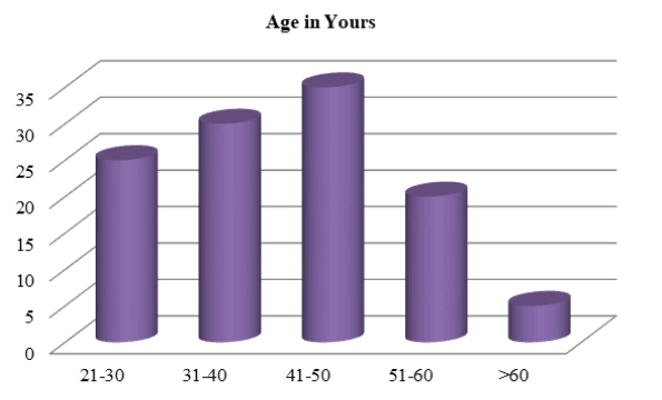
It is observed from the present study that majority 20 patients (31.7%) were in the age group 41 years to 50 years. The mean age in the present study group was (40.8) years with standard deviation of (11.5) years (FIGURE 1).
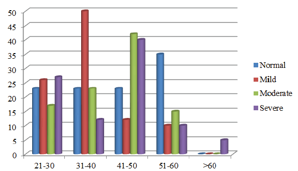
In the present study prevalence was highest in the 41-50 age group of which 2 (22.2%) were normal, 2 (12.5%) had mild dry eye, 10 (43.5%) had moderate dry eye, 6 (40%) had severe dry eye (FIGURE 2).
In the present study TBUT of patient studied in association of dry eye showed right eye mean of 9.5 ± 2 standard deviation in mild dry eye group, mean of 6.39 ± 2.35 standard deviation in moderate dry eye group, mean of 3.20 ± 1.15 standard deviation in the severe dry eye group showing a significant p value of less than 0.001. Whereas in the left eye mean of 10.38 ± 2.42 standard deviation in the mild dry eye group, mean of 7.09 ± 2.48 standard deviation in the moderate dry eye group, mean of 3.67 ± 1.05 standard deviation of in severe dry eye group showing a p value of less than 0.001 (TABLE 1).
| TBUT Test | Dry Eye | Total | P value | |||
|---|---|---|---|---|---|---|
| Normal | Mild | Moderate | Severe | |||
| Right Eye | 11.00 ± 2.45 | 9.50 ± 2.00 | 6.39 ± 2.35 | 3.20 ± 1.15 | 7.08 ± 3.40 | < 0.001** |
| Left Eye | 12.00 ± 2.96 | 10.38 ± 2.42 | 7.09 ± 2.48 | 3.67 ± 1.05 | 7.81 ± 3.68 | < 0.001** |
In the present study schirmer’s test of patient studied with the prevalence of in right eye mean of 10.19 ± 2.81 of standard deviation in the mild dry eye group, mean 8.26 with ± 3.40 of standard deviation in the moderate dry eye group, mean of 4.47 with ± 0.83 standard deviation in the severe dry eye group. For left eye mean of 10.88 with ± 3.16 of standard deviation in the mild dry eye group, mean of 8.91with ± 3.59 of standard deviation in the moderate dry eye group, mean of 4.47 ± 1.30 of standard deviation in the severe dry eye group with p value for both eyes being highly significant p< 0.001 (TABLE 2).
| Schirmer’s test | Dry Eye | Total | P value | |||
|---|---|---|---|---|---|---|
| Normal | Mild | Moderate | Severe | |||
| Right Eye | 12.33 ± 3.00 | 10.19 ± 2.81 | 8.26 ± 3.40 | 4.47 ± 0.83 | 8.43 ± 3.76 | < 0.001** |
| Left Eye | 13.33 ± 4.36 | 10.88 ± 3.16 | 8.91 ± 3.59 | 4.87 ± 1.30 | 9.08 ± 4.19 | < 0.001** |
In the present study Rose Bengal test of patient studied with the prevalence of in right eye mean of 4.19 ± 1.52 standard deviation in the mild dry eye group, mean of 5.22 with ± 1.31 standard deviation in the moderate dry eye group, mean of 7.47 with ± 0.83 standard deviation in the severe dry eye group. For left eye mean of 4.12 with ± 1.59 standard deviation in the mild dry eye group, mean of 5.22 with ± 1.53 of standard deviation in the moderate dry eye group, mean of 7.67 with ± 0.82 standard deviation in the severe dry eye group with p value for both eyes being highly significant p< 0.001 (ANNOVA TEST) (TABLE 3).
| Rose Bengal Test | Dry Eye | Total | P value | |||
|---|---|---|---|---|---|---|
| Normal | Mild | Moderate | Severe | |||
| Right Eye | 2.89 ± 1.05 | 4.19 ± 1.52 | 5.22 ± 1.31 | 7.47 ± 0.83 | 5.15 ± 1.94 | < 0.001** |
| Left Eye | 2.89 ± 1.16 | 4.12 ± 1.59 | 5.22 ± 1.53 | 7.67 ± 0.82 | 5.19 ± 2.07 | < 0.001** |
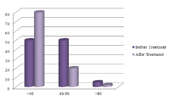
In the present study of assessing OSDI on scale of 0-100 shows scale of < 40 before treatment group as 27 (42.9%), after treatment 47 (74.6%), having a percentage change of 31.7%. In the scale of 40-80, 31 (49.2%) before treatment, 15 (23.8%) after treatment, having a percentage change of -25.4%. In the scale of > 80.5 (7.9%) before treatment group, 1 (1.6%) after treatment group showing a percentage change of -6.3%. The present study of patients before treatment in association with the prevalence of dry eye shows OSDI score of 41.10 mean with ± 23.84 standard deviation, on OSDI scale 41.90 with ± 23.51 standard deviation both showing p value less than (p< 0.001) highly significant (FIGURE 3).
In the present study prevalence of dry eye patients after treatment was associated with OSDI score mean of 29.08 with ± 18.78 standard deviation and scale showed mean 0f 29with ± 18.55 standard deviation both showing p value less than(p< 0.0001)highly significant (TABLE 4 and TABLE 5).
| Before treatment | Dry eye | Total | P value | |||
|---|---|---|---|---|---|---|
| Normal | Mild | Moderate | Severe | |||
| OSDI Score | 11.32 ± 4.49 | 23.79 ± 5.37 | 42.15 ± 9.63 | 75.81 ± 11.14 | 41.10 ± 23.84 | < 0.001** |
| OSDI Scale | 12.97 ± 1.55 | 25.04 ± 7.65 | 42.81 ± 10.46 | 75.85 ± 10.63 | 41.90 ± 23.51 | < 0.001** |
| After treatment | Dry eye | Total | P value | |||
|---|---|---|---|---|---|---|
| Normal | Mild | Moderate | Severe | |||
| OSDI Score | 10.86 ± 4.57 | 14.61 ± 4.07 | 28.24 ± 7.07 | 56.71 ± 13.49 | 29.08 ± 18.78 | < 0.001** |
| OSDI Scale | 12.97 ± 1.55 | 13.73 ± 5.66 | 28.17 ± 5.63 | 56.81 ± 13.31 | 29.15 ± 18.55 | < 0.001** |
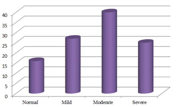
In the present study prevalence of dry eye was noted as follows mild dry eye 16 (25.4%), moderate dry eye 23 (36.5%), severe dry eye 15 (23.8%) (FIGURE 4).
In the present study correlation among the various test like TBUT, Schirmer’s, Rose Bengal test, OSDI showed good correlation with significant p value among all p< 0.001 highly significant. In our study comparison of variable according to the prevalence of dry eye with regards to the OSDI, TBUT, Schirmer’s, rose Bengal test reveals strongly significant p value. OSDI mean 41.10 ± 23.84 standard deviation, TBUT mean 7.44 ± 3.51 standard deviation, schirmer’s test mean 8.75 ± 3.92 standard deviation, Rose Bengal mean 5.17 ± 1.98 standard deviation overall validated the tests done.
Result
Majority 31.7% patients were in the group 41 years to 50 years. In the study OSDI questionnaire had a good reliability and consistency (p< 0.001) highly significant. Pearson correlation with r value among various test like Rose Bengal test, Schirmer’s test and TBUT showed a good correlation.
Discussion
The present study revealed that there is appreciable variation among diagnostic test results among different diseases and the best test combination to detect dry eye is OSDI/TBUT/Schirmer/Rose Bengal test. This result emphasises the importance of most commonly used tests to detect dry eye in clinical practice and also their variability. Meaningful diagnostic testing in dry eye disease across a broad range of different aetiologies and presentations is still a challenge [9]. Owing to great variability in dry eye disease severity, it is unlikely that a single test result has adequate sensitivity to serve for dry eye given its multifactorial nature and numerous manifestations. So that’s why research on potential dry eye diagnostic tools and therapeutic agents has increased exponentially. The correlation between subjective and objective findings was good but not in all cases and could be caused by multifactorial nature of dry eye syndrome. In our study 50 (79.4%) of patients have some symptoms such as foreign body sensation, burning, grittiness related to dry eye. there was good association between subjective symptoms of dry eye and its validation with objective test like, Tbut, Schirmer’s, Rose Bengal test [10], objective studies of dry eye commonly involved TBUT, Schirmer’s, Rose Bengal test (bjisterveldâ??s score) and when used in our study showed significant p value of less than 0.001. Also in the present study the OSDI demonstrated consistency and good test reliability. The OSDI also demonstrated excellent validity effectively discriminating between normal, mild, moderate and severe dry eye diseases as defined by both the physician assessment of severity and composite disease severity score [11]. Pearson correlation with r value shows perfect correlation between test and significant p value (0.001) OSDI score/scale showed strongly significant value with dry eye before and after treatment (p less than 0.001). OSDI was very well correlated with the clinical tests done suggesting that it is a very good tool if answered properly by the patients and can give the status of dry eye before moving to any tests.
Conclusions
Consensus regarding protocol in dealing with dry eye clinical diagnosis and standardisation of objective test is lacking. Dry eye is a complex multifactorial disease that involves the eyelids, tear film, ocular surface, immune and autonomic nervous system. The condition may be asymptomatic but often time causes significant patient discomfort and visual degradation. More studies into the association of risk factors in dry eye causation needs to be done. Finding an optimal treatment regimen can often be challenging and is a condition that requires patience from both the physician and patient. While mild case require annual visit, most patients suffering from moderate to severe dry eye require frequent monitoring for potential complications of dry eye and treatment efficacy. However the successful treatment of dry eye can lead to significant patient satisfaction and improved quality of life.
References
- Begley CG, Chalmers RL, Abetz L, et al. The Relationship between habitual patient reports symptoms and clinical signs among patients with dry eye of varying severity. Invest Ophthalmol Vi Sci. 44(11), 4753-4761(2003).
- Yanoff M, Duker JS. Ophthalmology. Elsevier. 3, 324-329 (2009).
[Google Scholar] [CrossRef]
- International dry eye workshop subcommittee. The definition and classification of dry eye disease. Ocul Surf. 5(2), 75-92 (2007).
- Plugfelder SC, Stephen C, Solomon, Stern, Michael. The diagnosis and management of dry eye 25 years review. Cornea. 19(5), 644-699 (2000).
[Google Scholar] [CrossRef]
- Afonso AA, Monroy D, Stern ME et al. Correlation of tear flourescein clearance and Schirmer test score with ocular irritations symptoms. Ophthalmology. 106(4), 803-810 (1999).
- Schaumberg DA, Buring JE, Sullivan DA, Dana MR. Hormone replacement therapy and dry eye syndrome. JAMA. 286(17), 2114-2119 (2001).
- Keith MS, Bartlett JD, Sudharshan L, Snedecor SJ. Associations between signs and symptoms of dry eye disease: a systematic review. Clin Ophthalmol. 9, 1719-1730 (2015).
- Mizuno Y, Yamada M, Miyake Y. Association between clinical diagnostic tests and health-related quality of life surveys in patients with dry eye syndrome. Jpn J Ophthalmol. 54(4), 259-265 (2010).
- Sullivan BD, Crews LA, Messmer EM, et al. Correllations between commonly used objective signs and symptoms for the diagnosis of dry eye disease: Clinical implications. Acta Opthalmol. 92(2), 161-166 (2014).
- Golding TR, Brennan NA. Diagnostics accuracy and intercorrelation of clinical tests for dry eye. Invest Ophthalmol Vis Sci. 34(4), 823 (1993).
[Google Scholar] [CrossRef]
- Grubbs JR, Tolleson-Rinehart S, Huynh K, Davis Rm. A review of quality of life measures in dry eye questionnaires. Cornea. 2014, 33(2), 215-218 (2014).
