Research Article - (2022) Volume 5, Issue 3
Prevalence of Lipid Abnormalities (Low HDL, High TG) Among Angiographically Proved CAD Patients
Md. Amir Hossain1*, Muhammed Akhtaruzzaman2, Monir Uddin Ahmed3, Mustafa Aolad Hossain4, Arman Ibne Huq5 and Md. Rezaul Karim62Associate Professor, Department of Cardiology, Bangladesh Medical College, Dhaka, Bangladesh
3Assistant Professor, Radiology & Imaging department, National Institute of Ophthalmology & Hospital , Bangladesh
4Resident Physician, Department of Medicine, Bangladesh Medical College Hospital, Dhaka, Bangladesh
5Assistant Professor, Department of Psychiatry, Bangladesh Medical College, Dhaka, Bangladesh
6Assistant Professor, Cardiology, Rajshahi Medical College, Rajshahi, Bangladesh
Received: Oct 27, 2022, Manuscript No. AMDHS-22-78321; Editor assigned: Oct 28, 2022, Pre QC No. AMDHS-22-78321 (PQ); Reviewed: Oct 30, 2022, QC No. AMDHS-22-78321 (Q); Revised: Oct 31, 2022, Manuscript No. AMDHS-22-78321 (R); Published: Nov 05, 2022, DOI: 10.5530/amdhs.2022.3.6
Abstract
Introduction: Dyslipidemia is a major primary risk factor for Coronary Artery Disease (CAD). Asians differs in prevalence of various lipid abnormalities than non-Asians. This study was conducted with the objective to assess the prevalence and pattern of lipid abnormalities and there correlation in patients with angiographically documented Coronary Artery Disease (CAD).
Objective: This study sought to determine the prevalence of lipid abnormality, specially in the form of low HDL-C and high TG, among patients with significant Coronary Artery Disease (CAD), (stenosis ≥ 50%) determined through coronary angiography as this is not well described in this population.
Methods: In a cross-sectional study, observational study, among 112 patients who underwent coronary angiography, we studied 81 cases who were diagnosed with significant CAD with stenosis ≥ 50%, as shown angiographically were found to have >50% coronary stenosis, defined as significant CAD. All patients were evaluated for risk factors and blood samples were collected for measuring fasting lipid profile in all patients during the admission for the coronary angiography. The data was analyzed to find statistical evidence for association between HDL-C and the pattern of CAD.
Results: Among 112 cases, 95 were diagnosed with significant CAD while 17 had non-significant disease. These 95 cases were include in the study. The mean age ( ± Standard deviation) was 53.63 ± 8.85, ranging from 28 years to 75 years. The majority of patients were males representing % of the cases {81/95 or 91/112}. The lipid profile analysis revealed that the mean total cholesterol was 179.79 ± 55.46 mg/dl, mean Low-Density Lipoprotein Cholesterol (LDL-C) was 103.54 ± 42.83 mg/dl, mean High Density Lipoprotein Cholesterol (HDL-C) was low, 34.47 ± 6.98 mg/dl and mean triglyceride level was high, 200.06 ± 143.3 mg/dl. 88% of patients had some form of dyslipidemia. The most frequent form of dyslipidemia among these patients was low levels of High-Density Lipoprotein Cholesterol (HDL-C) among 86.6% of the patients followed by 60.7% of patients with high Triglyceridemia (TG), 30.4% of the patients having high Total Cholesterol (TC) and 24.7% with high Low-Density Lipoprotein Cholesterol (LDL-C). 17 (15.2%) patients had normal coronary arteries, 32 (28.6%) had one vessel disease, 29 (25.9 %) had two vessel disease and 30 (26.8%) had three vessel disease.
Conclusion: Dyslipidemia is a highly prevalent risk factor among Bangladeshi patients with CAD. Low HDL-C was the most frequent lipid abnormality. Hypertriglyceridemia and low HDL cholesterol is common in patients with CAD compared with hypercholesterolemia and high LDL-C. This suggests the need to rethink treatment thresholds and targets in this population. The low HDL-C level among Bangladeshi patients require further study and targeted intervention.
Keywords
Prevalence, Lipid abnormalities, Angiographically, CAD
Introduction
According to WHO, Cardiovascular Diseases (CVD) are by far the largest cause of death worldwide, representing 31% of the annual deaths globally in 2015. Like elsewhere, CVDs are also a huge burden for Bangladesh and accounts for 17% of the annual deaths (World Health OrganizationNoncommunicable Diseases (NCD) Country Profiles, 2014).Coronary Artery Disease (CAD), one of the CVDs, is an important medical and public health issue currently, because it is one of the most common and leading causes of death [1]. Bangladesh has been experiencing an epidemiological transition from communicable to Non-Communicable Diseases (NCD) and hence CAD poses as an emerging threat. Conventional cardiovascular risk factors, such as hypertension, diabetes, smoking, and dyslipidemia, increase the risk of developing Coronary Artery Disease (CAD) [2-4]. Coronary Artery Disease (CAD) is defined as the occlusion of coronary arteries due to atherosclerosis which leads to impairment of blood supply to the heart. Depending on the severity of this stenosis it may lead to various outcomes. Atherosclerosis is defined as the thickening of arteries due to formation of plaques within the inner layer of the artery due to deposition of excess fat, macrophages, fibrous tissue and other cellular debris often occluding the vessels. As described by the pathology, dyslipidemia is one of the major contributors of atherosclerosis. Although at different times, different lipoproteins have been associated with atherosclerosis, contrary to the previous believes as LDL being the predominant lipoprotein contributing to CAD, a number of epidemiological studies have portrayed a strong association between low levels of high density lipoprotein (HDL) cholesterol and development and progression of atherosclerotic Coronary Artery Disease. Primary prevention studies have shown that the early detection and aggressive treatment of risk factors prevent cardiovascular events [5, 6]. One of the largest of such studies, the INTERHEART study, was a worldwide study designed to assess the relevance of different risk factors on myocardial infarct development, 85% had at least one of the conventional risk factors [7, 8]. Knowledge of a baseline lipid level may help in therapeutic decisions of initiation of lipid-lowering treatment and the patient’s willingness to adhere to a recommendation for long-term lipid-lowering therapy. Previous reports of the prevalence of the risk factors and lipid profiles in CAD have been done in patients without considering the presence or absence of coronary lesions. The aims of our study were to investigate the prevalence of abnormal lipid profiles at the time of admission in a cohort of patients with and significant CAD (stenosis ≥ 50%) determined through coronary angiography.
Materials and Methods
This was a retrospective cross-sectional study. We studied the pattern and prevalence of dyslipidemia and sought to find the most common lipid abnormalities among angiographically proved CAD patients. Information was gathered from medical records of patients admitted for coronary angiography between 2018 and 2019 in a Tertiary Hospital, Dhaka, Bangladesh. Coronary angiography was performed in the catheterization laboratory of the institute and interpreted by interventionist cardiologists. Reporting was done regarding the stenosis percentage of the main epicardial coronary arteries, and the extent of CAD was categorized as one-vessel, two-vessel, or three-vessel disease, according to the number of affected vessels. Only patients with significant CAD, defined as the presence of ≥ 50% stenosis of any of the epicardial vessels, were included in the study. Patients with normal coronary angiography or mild disease, defined as <50% stenosis in any of the epicardial vessels, were excluded.
Lipid profile measurements were done routinely, within 24 hours of admission of the patients, in fasting state. All results were obtained from the same pathological laboratory (i.e. within the hospital) and thus ensured maintenance strategy and standardization. Lipid profile parameters namely Total Cholesterol (TC), TG, HDL-C/HDL, LDL-C/LDL were recorded as variables. Lipid profile evaluation is done in this laboratory by end point kinetic method by Roche, for TC, TG and HDL-C, while LDL-C is calculated with the Friedewald formula unless TG are elevated (greater than 400 mg/dL): Friedewald formula, in mg/dL:
• LDL-C ¼ TC - HDL-C - TG/5.
• Dyslipidemia was defined as high serum levels of TC ≥ 200 mg/dL, LDL-C ≥ 130 mg/dL, and TG ≥ 150 mg/dL; a low serum level of HDL-C was defined as ≤ 40 mg/dL [9].
Statistical Analysis
Statistical analysis were performed with SPSS version 20 (SPSS, Inc., Chicago, IL, USA) statistical software. Categorical variables were reported by frequency and percentage; groups were compared using the chi-square test. Continuous variables were reported as means and standard deviation. All p-values were reported as 2-tailed with statistical significance set at 0.05 and confidence intervals calculated at the 95%.
Results
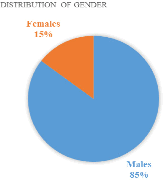
During the study period, we identified 112 patients who were subjected to coronary angiography. 17 patients were excluded due to normal coronary results and/or nonsignificant lesions. We thus analyzed 95 patients with significant coronary stenosis, ≥ 50%. The mean age of patients were 54.40 years (± SD 8.78 years), where the minimum age was 28 and maximum age was 75. Majority of the patients were young, with around 78.9% (75) of the patients being less than or equal to 60 years of age. The study consisted predominantly of males as represented by 85.3% of the patients, while 14.7% were females (81 versus 14), as shown in FIGURE 1.
Figure 1: Distribution of gender
The lipid profile analysis revealed that the mean total cholesterol was 182.60 ± 54.41 mg/dl, mean Low-Density Lipoprotein Cholesterol (LDL-C) was 105.46 ± 40.85 mg/dL, mean High Density Lipoprotein Cholesterol (HDL-C) was low, 34.26 ± 7.234 mg/dL and mean triglyceride level was high, 207.64 ± 151.20 mg/dL. The mean levels of different lipids are shown in TABLE 1.
| Lipid parameters | N | Minimum | Maximum | Mean | Std. Deviation |
|---|---|---|---|---|---|
|
Total Cholesterol (mg/dL) |
95 | 80 | 482 | 182.6 | 54.413 |
|
HDL (mg/dL) |
95 | 21 | 64 | 34.26 | 7.234 |
|
Triglyceride (mg/dL) |
95 | 48 | 948 | 207.64 | 151.2 |
|
LDL (mg/dL) |
95 | 14 | 198 | 105.46 | 40.85 |
The mean levels of the different lipid parameters were quite similar between the two categories of gender, with only significant difference (<0.05) in mean total cholesterol among males and females. Means of TG, LDL-C and HDL-C were equally high, normal and low respectively (TABLE 2).
| Lipid levels among genders | Gender of Patient | N | Mean | SD |
|---|---|---|---|---|
| Total Cholesterol (mg/dL) | Males | 81 | 177.52 | 53.45 |
| Females | 14 | 212 | 45.12 | |
| LDL (mg/dL) | Males | 81 | 105.1 | 43.25 |
| Females | 14 | 107.57 | 35.26 | |
| HDL (mg/dL) | Males | 81 | 34.07 | 41.7 |
| Females | 14 | 35.36 | 36.26 | |
| Triglyceride (mg/dL) | Males | 81 | 208.79 | 110.99 |
| Females | 14 | 201 | 102.45 |
In this study, the most frequent form of dyslipidemia among the patients with significant CAD was found to be low levels of HDL-C (<40mg/dL)at 86.3%, followed by high TG levels (≥ 150mg/dL) to be 63.2%, high levels of total cholesterol (≥ 200mg/dL) at 31.6%, and high LDL-C (≥ 130mg/dL) to be 27.4% (TABLE 3).
| Proportion of dyslipidemia | Number of patients (N) | Percentage (%) |
|---|---|---|
| High TC | 30 | 31.60% |
| Low HDL-C | 82 | 86.30% |
| High LDL-C | 26 | 27.40% |
| High TG â?? | 60 | 63.20% |
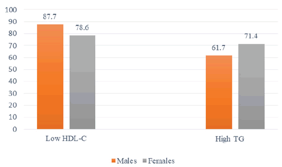
The males had a higher tendency of having low HDL than females 87.7% vs 78.6%, while females were more frequently having high TG than their male counterparts, 71.4% vs 61.7% shown in the bar chart in FIGURE 2. A significant co-relation (<0.05, r=0.2) was found between High TG and low HDL levels.
Figure 2: Between high TG and low HDL levels.
References
- Islam AM, Majumder AA. Coronary artery disease in Bangladesh: A review. Indian heart j. 65(4), 424-435 (2013).
- González-Pacheco H, Vargas-Barrón J, Vallejo M et al. Prevalence of conventional risk factors and lipid profiles in patients with acute coronary syndrome and significant coronary disease. Ther clin risk manag. 10, 815 (2014)
- Grundy SM, Pasternak R, Greenland P, Smith S, Fuster V. Assessment of cardiovascular risk by use of multiple-risk-factor assessment equations: A statement for healthcare professionals from the American Heart Association and the American College of Cardiology. Circulation. 100(13), 1481-1492 (1999).
[Google Scholar] [CrossRef]
- Pasternak RC, Grundy SM, Levy D, Thompson PD. Task Force 3. Spectrum of risk factors for coronary heart disease. J Am Coll Cardiol. 27(5), 978-990 (1996).
- Heart Protection Study Collaborative Group. MRC/BHF Heart Protection Study of cholesterol lowering with simvastatin in 20,536 high-risk individuals: a randomised placebo-controlled trial. Lancet. 360(9326), 7-22 (2002). [Google Scholar]
[CrossRef]
- Sever PS, Dahlöf B, Poulter NR et al. Prevention of coronary and stroke events with atorvastatin in hypertensive patients who have average or lower-than-average cholesterol concentrations, in the Anglo-Scandinavian Cardiac Outcomes Trial – Lipid Lowering Arm (ASCOT-LLA): a multicentre randomized controlled trial. Lancet. 361(9364), 1149-1158 (2003).
- Yusuf S, Hawken S, Ounpuu S et al. Effect of potentially modifiable risk factors associated with myocardial infarction in 52 countries (the INTERHEART study): case-control study. Lancet. 364(9438), 937-952 (2004).
[Google Scholar] [CrossRef]
- Khot UN, Khot MB, Bajzer CT et al. Prevalence of conventional risk factors in patients with coronary heart disease. JAMA. 290(7), 898 (2003).
- Cannon CP, Battler A, Brindis RG et al. American College of Cardiology key data elements and definitions for measuring the clinical management and outcomes of patients with acute coronary syndromes: a report of the American College of Cardiology Task Force on clinical data standards (acute coronary syndromes writing committee) endorsed by the American Association of Cardiovascular and Pulmonary Rehabilitation, American College of Emergency Physicians, American Heart Association, Cardiac Society of Australia & New Zealand, National Heart Foundation of Australia, Society for Cardiac Angiography and Interventions, and the Taiwan Society of Cardiology. J Am Coll Cardiol. 38(7), 2114-2130 (2001).
[Google Scholar] [CrossRef]
- Dey S, Flather MD, Devlin G et al. Sex-related differences in the presentation, treatment and outcomes among patients with acute coronary syndromes: the Global Registry of Acute Coronary Events. Heart. 95(1), 20-26 (2009).
- Silbiger JJ, Ashtiani R, Attari M et al. Atheroscerlotic heart disease in Bangladeshi immigrants: Risk factors and angiographic findings. Int j cardiol. 146(2), e38-40 (2011).
- Gupta M, Brister S, Verma S. Is South Asian ethnicity an independent cardiovascular risk factor?. Can J Cardiol. 22(3), 193-197 (2006).
- Gupta M, Singh N, Verma S. South Asians and cardiovascular risk: what clinicians should know. Circulation. 113(25), e924-e992 (2006).
- Andreotti F, Marchese N. Women and coronary disease. Heart. 94(1), 108-116 (2008).
- Boden WE, O’Rourke RA, Teo KK et al. The evolving pattern of symptomatic coronary artery disease in the United States and Canada: baseline characteristics of the Clinical Outcomes Utilizing Revascularization and Aggressive DruG Evaluation (COURAGE) trial. Am j cardiol. 99(2), 208-212 (2007).
- Assmann G, Schulte H, von Eckardstein A, Huang Y. High-density lipoprotein cholesterol as a predictor of coronary heart disease risk. The PROCAM experience and pathophysiological implications for reverse cholesterol transport. Atherosclerosis. 124, S11-S20 (1996).
- Cooney MT, Dudina A, De Bacquer D et al. HDL cholesterol protects against cardiovascular disease in both genders, at all ages and at all levels of risk. Atherosclerosis. 206, 611-616 (2009).
- Maron DJ. The epidemiology of low levels of high-density lipoprotein cholesterol in patients with and without coronary artery disease. Am j cardiol. 86(12), 11-14 (2000).
- Gupta R, Gupta VP, Sarna M et al. Prevalence of coronary heart disease and risk factors in an urban Indian population: Jaipur Heart Watch-2. Indian heart j. 54(1), 59-66 (2002).
[Google Scholar] [CrossRef]
- Goel PK, Bharti BB, Pandey CM et al. A tertiary care hospital-based study of conventional risk factors including lipid profile in proven coronary artery disease. Indian heart j. 55, 234-240 (2003).
[Google Scholar] [CrossRef]
- Mohan V, Deepa R, Rani SS et al. Prevalence of coronary artery disease and its relationship to lipid in a selected population in South India. J Am Coll Cardiol. 38, 682-687 (2001).
[Google Scholar] [CrossRef]
- Chapman MJ, Ginsberg HN, Amarenco P et al. Triglyceride-rich lipoprotein and high-density lipoprotein cholesterol in patients at high risk of cardiovascular disease: evidence and guidance for management. Eur Heart J. 32(11), 1345-1361 (2011).
- Reddy KS, Prabhakaran D, Chaturvedi V et al. Methods for establishing a surveillance system for cardiovascular diseases in Indian industrial populations. Bull World Health Organ. 84, 461-467 (2006).
[Google Scholar] [CrossRef]
- Ibrahim MM, Ibrahim A, Shaheen K, Nour MA. Lipid profile in Egyptian patients with coronary artery disease. Egypt Heart J. 65(2), 79-85 (2013).
- Sharma SB, Garg S, Veerwal A, Dwivedi S. hs-CRP and oxidative stress in young CAD patients: A pilot study. Indian J Clin Biochem. 23, 334-336 (2008).
- Mohan V, Ramachandran A, Snehalatha C et al. High prevalence of diabetes and impaired glucose tolerance in India: National Urban Diabetes Survey. Diabetologia. 44, 1094-1101 (2001).


